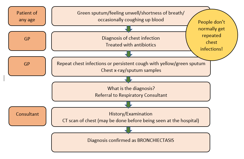
Bronchiectasis is diagnosed by a CT scan of the chest. A normal chest x-ray cannot confirm bronchiectasis – nor can it be used to say that bronchiectasis is not present. Most patients have had a CT of their chest requested because of chronic symptoms in keeping with bronchiectasis. At this point patients are reviewed by the respiratory team in secondary care who will arrange for further investigations such as breathing tests, blood tests and sputum culture.
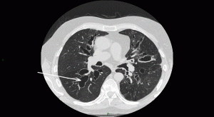
The CT scan shows airways that are permanently damaged and look like cysts. Due to the appearance of the airways, this type of bronchiectasis is described as cystic* bronchiectasis. An arrow points to the these cysts. The changes seen here are quite severe.
* In this context the term cystic is not related to cystic fibrosis. It is purely used to describe the shape of the airways (typically this can be associated with more moderate to severe bronchiectasis).
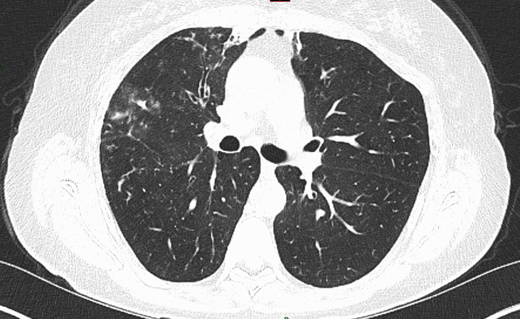
This CT image shows lungs with milder bronchiectasis. There is a wide range of changes that can be seen in a CT scan of the chest, with mild bronchiectasis through to more severe changes.